 Structural alterations in chromosomes.
Breakage of a chromosome can lead to four types of changes in chromosome structure.
- Deletion
- Duplication
- Inversion
- Translocation
A deletion occurs when a chromosomal fragment lacking a centromere is lost. The affected chromosome is then missing certain genes. In some cases, if meiosis is in progress, such a "deleted" fragment may become attached as an extra segment to a sister chromatid, producing a duplication. Alternatively, a detached fragment could attach to a non–sister chromatid of a homologous chromosome. In that case, the "duplicated" segments might not be identical because the homologues could carry different alleles of certain genes. A chromosomal fragment may also reattach to the original chromosome but in the reverse orientation, producing an inversion. A fourth possible result of chromosomal breakage is for the fragment to join a non–homologous chromosome, a rearrangement called a translocation. Deletions and duplications are especially likely to occur during meiosis.
In crossing over, non–sister chromatids sometimes break and rejoin at "incorrect" places, so that one partner gives up more genes than it receives. The products of such a nonreciprocal crossover are one chromosome with a deletion and one chromosome with a duplication. A diploid embryo that is homozygous for a large deletion (or has a single X chromosome with a large deletion, in a male) is usually missing a number of essential genes, a condition that is ordinarily lethal. Duplications and translocations also tend to have harmful effects. In reciprocal translocations, in which segments are exchanged between non homologous chromosomes, and in inversions, the balance of genes is not abnormal – all genes are present in their normal doses. Nevertheless, translocations and inversions can alter phenotype because a gene's expression can be influenced by its location among neighboring genes.
") Karyotype of Down syndrome (Trisomy 21)
Karyotype of Down syndrome (Trisomy 21)
Down's syndrome is a chromosomal abnormality resulting in mental handicap and a characteristic physical appearance. Down syndrome was described for the first time by Langdon Down in 1866. The affected individuals were observed to have a different and characteristic appearance. Down syndrome, affects approximately one out of every 700 children born in the United States.
Down syndrome is usually the result of aneuploidy, an extra chromosome 21, so that each body cell has a total of 47 chromosomes. Because the cells are trisomic for chromosome 21, Down syndrome is often called trisomy 21.
Down syndrome includes characteristic facial features, short stature, heart defects, susceptibility to respiratory infection, and mental retardation. Furthermore, individuals with Down syndrome are prone to developing leukemia and Alzheimer's disease. Children with Down's syndrome have an I.Q. of between 30 and 80 (Normal range – 70 to 130).
Although people with Down syndrome, on average, have a life span shorter than normal, some live to middle age or beyond. Most are sexually underdeveloped and sterile.
 Diagnosing trisomy 21
The frequency of Down syndrome increases with the age of the mother. While the disorder occurs in just 0.04% of children born to women under age 30, the risk climbs to 1.25% for mothers in their early 30s and is even higher for older mothers. Because of this relatively high risk, pregnant women over 35 are candidates for fetal testing to check for trisomy 21 in the embryo.
The correlation of Down syndrome with maternal age has not yet been explained. Most cases result from non disjunction during meiosis I, and some research points to an age–dependent abnormality in a meiosis checkpoint that normally delays anaphase until all the kinetochores are attached to the spindle (like the M phase checkpoint of the mitotic cell cycle) .
Trisomies of some other chromosomes also increase in incidence with maternal age, although infants with these autosomal trisomies rarely survive for long. Today, Down syndrome is routinely identified in karyotypes or by using fluorescent probes for chromosome 21 through fluorescence in situ hybridization (FISH).
 Aneuploidy
Aneuploidy is an abnormal number of chromosomes, and is a type of chromosome abnormality. An extra or missing chromosome is a common cause of genetic disorders (birth defects). Aneuploidy occurs during cell division when the chromosomes do not separate properly between the two cells.
Chromosome abnormalities occur in 1 of 160 live births. Most cases of aneuploidy result in termination of the developing fetus, but there can be cases of live birth; the most common extra chromosomes among live births are 21, 18 and 13. Different species have different numbers of normal chromosomes and thus the term "aneuploidy" refers to the chromosome number being different for that species.
Aneuploidies disturb the delicate balance of gene products in cells. Because each chromosome contains hundreds of genes, the addition or loss of even a single chromosome disrupts the existing equilibrium in cells, and in most cases, is not compatible with life. Using the tools of modern cytogenetics, scientists have recently provided new insights into the origins of aneuploidy.
Researchers now appreciate that aneuploid gametes are produced at surprisingly high rates in human meiosis, and that very few aneuploid embryos are able to survive. Much attention is currently focused on determining how specific imbalances in gene expression lead to the profound phenotypes associated with aneuploid conditions, such as Down syndrome, with the ultimate goal of developing therapeutic interventions.
 Cri du Chat syndrome
Many deletions in human chromosomes, even in a heterozygous state, cause severe problems. One such syndrome, known as Cri du chat ("cry of the cat"), results from specific deletion in chromosome 5. Cri du chat syndrome, also known as Lejeune’s syndrome, is a rare genetic disorder due to a missing part of chromosome 5.
The syndrome gets its name from the characteristic cry of affected infants, which is similar to that of a meowing kitten, due to problems with the larynx and nervous system. About 1/3 of children lose the cry by age 2. A child born with this deletion is mentally retarded, has a small head with unusual facial features, and has a cry that sounds like the mewing of a distressed cat. Such individuals usually die in infancy or early childhood. Another type of chromosomal structural alteration associated with human disorders is translocation, the attachment of a fragment from one chromosome to another, non–homologous chromosome.
Chromosomal translocations have been implicated in certain cancers; including chronic myelogenous leukemia (CML) (Chronic myelogenous leukemia is cancer that starts inside bone marrow, the soft tissue inside bones that helps form blood cells. The cancer grows from cells that produce white blood cells). Leukemia is a cancer affecting the cells that give rise to white blood cells, and in the cancerous cells of CML patients, a reciprocal translocation has occurred.
In these cells, the exchange of a large portion chromosome 22 with a small fragment from a tip of chromosome 9 produces a much shortened, easily recognized chromosome 22, called the Philadelphia chromosome. (Philadelphia chromosome or Philadelphia translocation is a specific chromosomal abnormality that is associated with chronic myelogenous leukemia (CML). It is the result of a reciprocal translocation between chromosome 9 and 22).
 Extra X chromosome
This is the chromosomal disorder affects male physical and cognitive development. This is due to the presence of an extra copy of X-chromosome that results into a karyotype of 47, XXY. 47, XXY (or XXY) is a genetic condition caused when someone has two X chromosomes and one Y chromosome. Males normally have an X chromosome and a Y chromosome (46, XY), and females normally have two X chromosomes (46, XX). XXY is one of the most common genetic conditions, affecting about 1 in 660 genetic males. Klinefelter syndrome is named for Dr. Harry Klinefelter, who first reported its symptoms in 1942.
People with an XXY chromosomal arrangement have a Y chromosome, they are considered as genetic males. Most XXY individuals develop as males, often not knowing they have an extra chromosome. Some will develop the varied and often subtle characteristics associated with Klinefelter syndrome. Similar conditions are caused by additional X chromosomes (48, XXXY; 49, XXXXY), but they are much more rare. The more X chromosomes a person has, the stronger the physical characteristics and health problems tend to be, including intellectual disability. Such an individual has overall masculine development, however, the feminine development (development of breast i.e; Gynaecomastia) is also expressed. Such individuals are sterile.
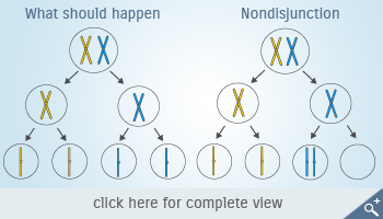 Nondisjunction
This is caused due to the phenomenon called nondisjunction. Nondisjunction is the failure of homologous chromosomes or sister chromatids to separate properly during cell division. This happens when a pair of sex chromosomes fails to separate during egg (or sperm) formation. When an egg or sperm with an extra X chromosomes joins with a normal sperm (or egg), the resulting embryo will end up with the sex chromosomes (XXY) instead of the normal two (XX or XY). Klinefelter syndrome and its variants are not inherited; these chromosomal changes usually occur as random events during the formation of reproductive cells (eggs and sperm) in a parent.
The chromosomal pattern- 47, XXY can be diagnosed using karyotype. A karyotype is an analysis of a patient's chromosomes taken from a blood sample. In some rare cases, this genetic disorder can be diagnosed during a woman's pregnancy. Doctors look for chromosome abnormalities in cells taken from the amniotic fluid surrounding the fetus (amniocentesis) or from the placenta (chorionic villus sampling, or CVS). Almost 10 percent of cases are diagnosed this way.